


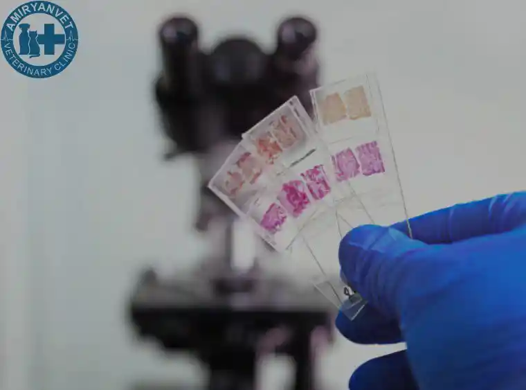
Microscopic examination – a type of laboratory test that uses a microscope to study the structure and properties of objects that are invisible to the naked eye. It is widely used in medical, veterinary, biological, and other scientific fields to detect microorganisms, cells, tissues, and to identify ...
Microscopic examination – a type of laboratory test that uses a microscope to study the structure and properties of objects that are invisible to the naked eye. It is widely used in medical, veterinary, biological, and other scientific fields to detect microorganisms, cells, tissues, and to identify the causes of various diseases.
Such an examination allows for the detection of infectious agents (e.g., microbes, viruses, or tuberculosis bacteria), cellular changes, or macro- and microscopic particles that may have diagnostic significance.
Through microscopic observations in veterinary medicine, many features of microbes can be determined:
Read more